What is a Sarcoma?
 A sarcoma is a malignant tumor and a rare type of cancer that occurs in adults, children and teens. Sarcomas comprise a complex family of cancers that cover a wide variety of distinct diseases that are categorized into two broad areas: soft tissues and bone. To learn more about the different types of sarcoma and to find out about our cancer treatment options, get in touch with the team at the Sarcoma Oncology Center in Santa Monica by calling 310-552-9999 or filling out our online contact form.
Soft Tissue Sarcomas
Soft tissue sarcomas are malignant tumors that manifest in the connective tissue of the tendon, ligament or muscle and are the most frequently occurring sarcomas. There are more than 75 different subtypes of soft tissue sarcomas. These subtypes differ in terms of their tissue of origin, genetic alterations, clinical behavior or sensitivity to certain therapies, age of onset, aggressiveness, and growth pattern.
Symptoms
Pain is the most common sarcoma symptom, as well as swelling and tenderness (from a tumor in or near a joint) or difficulty with normal movement. Other symptoms may include fatigue, fever, weight loss and anemia. Additionally, the presence of a painless lump which could eventually become sore or painful is something that an oncologist should check.
Anyone who is suffering from apparent sarcoma symptoms must consult with their physician and not assume they have cancer, as these symptoms are general and could be attributed to another medical condition.
Bone Sarcomas
Bone sarcomas are rare forms of cancer that form in the bone (including upper arm, shoulder, ribs and legs). There are four types of primary bone cancer: osteosarcomas, chondrosarcomas, the Ewing’s family of tumors, and giant cell tumor of the bone. Osteosarcoma and Ewing’s sarcoma occur more frequently in children and adolescents; chondrosarcoma and giant cell tumor of the bone occur more often in adults.
Since both soft tissue and bone sarcomas are rare, they appear in a small percentage of the juvenile and adult populations. While only 1% of adult cancers are diagnosed as soft tissue, approximately 15% of childhood cancers are soft tissue sarcoma. Bone sarcoma is less common with approximately 2,500 new cases each year for adults and children.
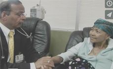
The oncologists at the Sarcoma Oncology Center are intimately involved in each patient’s care. We collaborate with radiologists, surgeons, pathologists and radiation oncologists at other prominent institutions to ensure that the complexity of each patient’s case is addressed in its entirety.
For more information on cancer treatment, visit WebMD.com.
Symptoms
Sarcoma symptoms can include the presence of a painless lump which could eventually become sore or painful, as well as blockage in the gastrointestinal system, or blood in vomit or stool.
Risk Factors
No specific attributable factors can be linked to cause soft tissue or bone sarcomas. However, there are a few factors that could increase the chances of developing soft tissue sarcomas:
- Exposure to ionizing radiation
- Exposure to certain chemicals (such as arsenic, vinyl chloride, herbicides containing phenoxyacetic acids, and wood preservatives containing chlorophenols)
- Family history (presence of inherited diseases including Gardner’s syndrome, neurofibromatosis, or Li-Fraumeni syndrome)
- Exposure to Agent Orange
Additionally, radiation treatments may also be a possible influencing factor for the risk of developing bone sarcoma.
Anyone who believes they manifest these symptoms must consult with their physician and not assume they have cancer as these symptoms are general and could be attributed to another medical condition.
Treatment and Medical Care
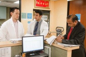 Sarcomas are a complex form of cancer and require team-oriented, multi-modal, individualized treatment plans. The Sarcoma Oncology Center in Santa Monica collaborates with prominent surgeons, pathologists and radiation oncologists from institutions such as UCLA, USC, Stanford, M.D. Anderson, St. John’s and Cedars Sinai to develop and implement the most effective multi-modal treatment plan for each case. The oncologists at the Sarcoma Oncology Center are intimately involved in each patient’s care to ensure that the complexity of each case is addressed in its entirety.
We develop treatment plans based on the grade and stage of a tumor, prior treatments received (if any), cancer progress, biopsy analysis and other individualized factors. The oncologists at the Sarcoma Oncology Center are at the forefront of sarcoma research and are pioneers of the most advanced sarcoma clinical research trials in the world. The oncologists at the Center in Santa Monica go beyond standard care for cancer when needed and have access to the most novel cancer treatment drugs at their fingertips.
Chemotherapy
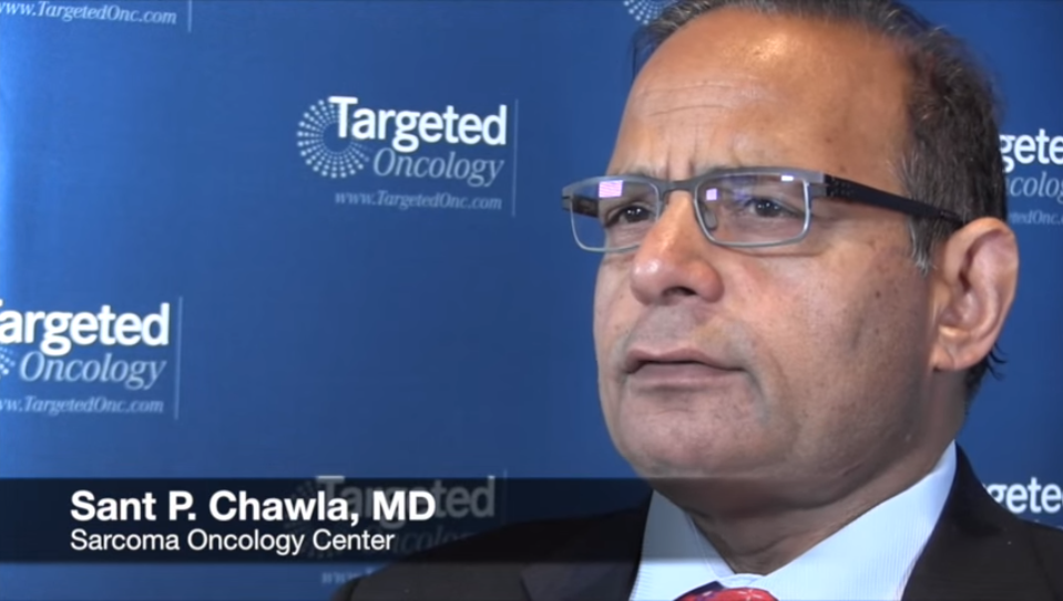
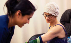
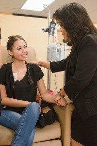
Chemotherapy is the most important treatment available for sarcomas because it is the only form of treatment which prevents and controls the spread of the cancer into the lungs, vital organs and other parts of the body. In the past, chemotherapy was highly toxic and had significant side effects. Thanks to new treatments pioneered by Dr. Chawla in collaboration with internationally renowned oncologists, the side effects of chemotherapy have been significantly reduced and are virtually nonexistent in many cases. New treatments protect the heart and kidney, substantially reduce infections and the need for blood transfusions and virtually eliminate nausea and vomiting for most patients. Chemotherapy is an art and science and the right combinations of treatments aggressively eliminates cancer, prevents recurrence and maintains the patient’s quality of life through treatment. Over 50% of Dr. Chawla’s patients can work during chemotherapy and maintain their daily routine activities and exercise through their treatment.
Surgery and Radiation
Surgery can be a particularly effective option after initial chemotherapy to shrink sarcoma tumors. With the advent of new adjuvant chemotherapy, improved technique in surgery and the availability of internal prostheses, amputation is now avoided in approximately 95% of sarcoma cases.
Radiation is another effective tool used to combat sarcomas in specific, localized areas. Radiation works by sterilizing the tumor cells and making them unable to divide and continue to grow.
Active Patient Participation
We encourage patients to participate actively in their medical care. When diagnosed with a sarcoma or malignant tumor, patients would want to consider discussing the following with their primary care physician and/or oncologist:
- What type of sarcoma do I have?
- Where is my sarcoma located? Has it spread from an initial site?
- What stage is my sarcoma?
- What are the treatment options?
- What is the specific course of treatment and why was it selected?
- What can I do to complete the course of treatment successfully and shorten recovery time?
- Are there clinical trials available?
In preparation for the initial appointment, we suggest patients write down their questions so that important questions and concerns are not omitted when meeting with the doctor.
Contact our Soft Tissue Cancer Treatment Specialists Today
Regardless of treatment approach, Sarcoma Oncology Center in Santa Monica offers patients the best possible solution and considers the physical and emotional well being of each individual. Please contact our center in Santa Monica at 310-552-9999 to schedule a consultation with one of our specialists in regards to malignant tumor treatment.
Next, learn more about cancer treatment.